Anti-PEG antibody ELISA (mouse IgG specific)
€ Preis anfragen
Polyethylene glycol (PEG) chains are often used to modify therapeutic biologic agents in order to prolong the circulating half-life of the modified protein. It has been reported that repeat injections of PEGylated proteins can induce anti-PEG antibodies. This assay employs the sandwich enzyme immunoassay technique. Immobilized PEG is coated onto a 96 well microplate. Calibrator and test samples are pipetted into the appropriate wells. Anti-PEG antibodies present in biological matrices is bound by the immobilized PEG. After washing away any unbound substances, enzyme linked anti IgG or IgM antibody is added to the wells. The plate is washed to remove any unbound antibody-enzyme reagent and a substrate solution is added to the wells for color development. The color development is proportional to the amount of anti-PEG antibodies present in test samples. The color development is stopped and the intensity of the color is measured
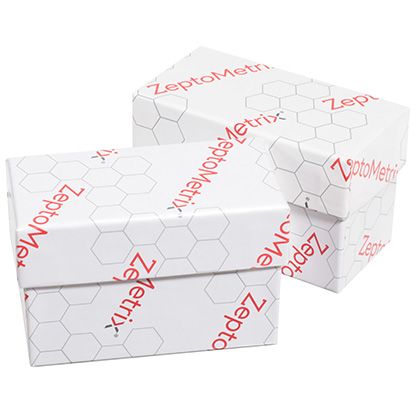
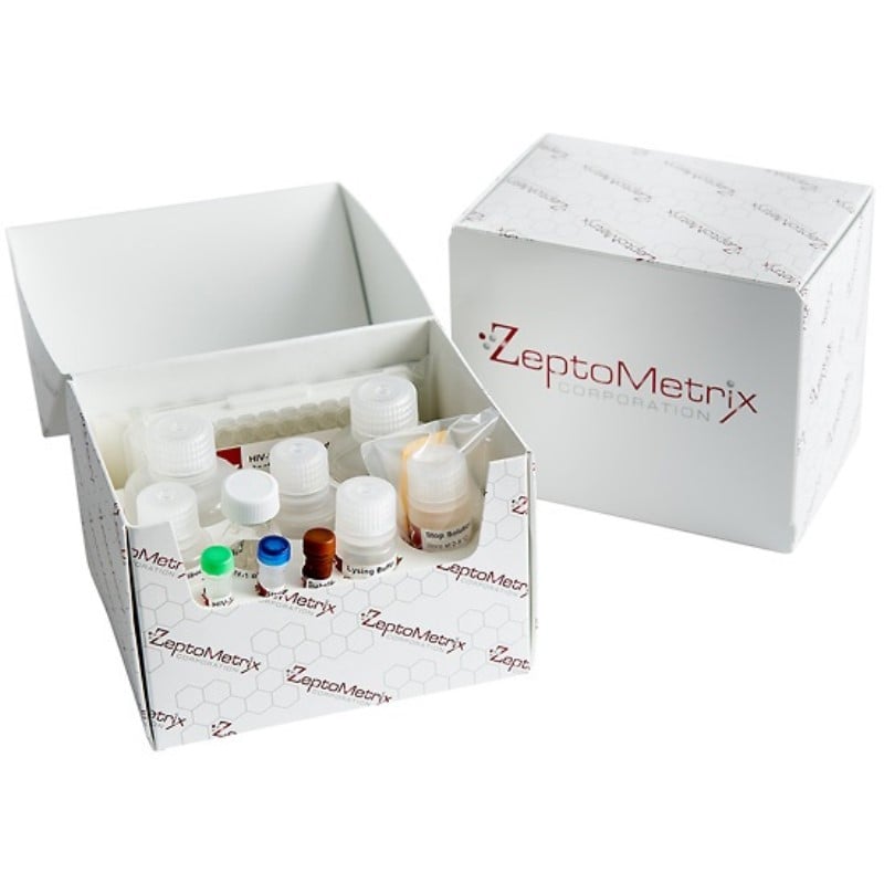
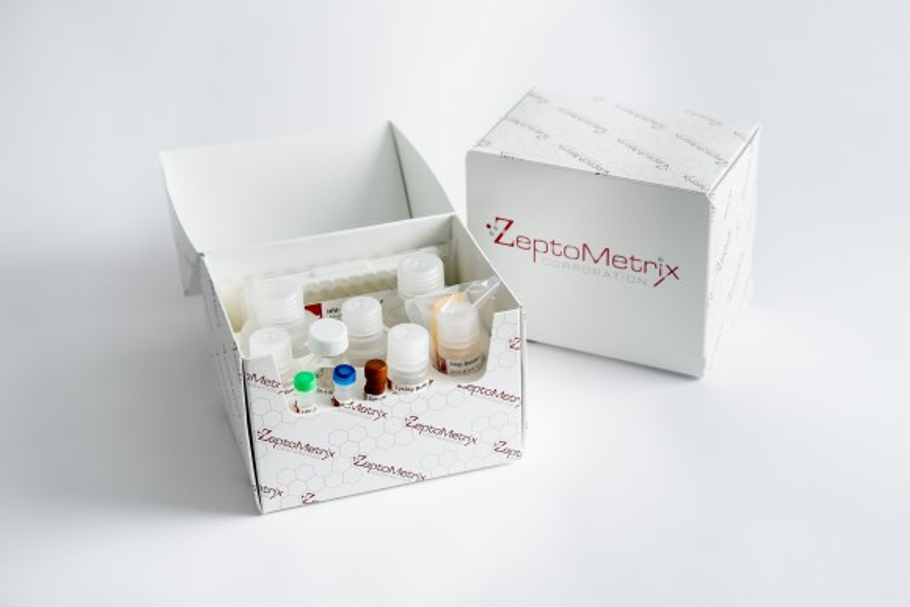
- Components: Coated microtiter plate, 96 wellsQC samples - 6x250ul10X wash buffer - 50 mlAssay buffer - 50ml1000X detection reagent - 17ulTMB - 12mlTMB stop solution - 12mlPlate sealers - 3
- Assay procedure: This assay employs the sandwich enzyme immunoassay technique. Immobilized PEG is coated onto a 96 well microplate. Calibrator and test samples are pipetted into the appropriate wells. Anti-PEG antibodies present in biological matrices is bound by the immobilized PEG. After washing away any unbound substances, enzyme linked anti IgG or IgM antibody is added to the wells. The plate is washed to remove any unbound antibody-enzyme reagent and a substrate solution is added to the wells for color development. The color development is proportional to the amount of anti-PEG antibodies present in test samples. The color development is stopped and the intensity of the color is measured
- Reagent preparation: Prepare only the appropriate amount of required reagent on the day of use. Store all reagents as per instructions stated on the label. 1. Wash Buffer (1X) Preparation Dilute wash buffer concentrate with deionized water 1/10 before use (for example add 20mL concentrate to 180mL deionized water). Mix well. 2. Detection Reagent (1X) Preparation: Dilute detection reagent with assay buffer 1/1000 before use (for example add 11μl concentrate to 11ml of assay buffer). Mix well. The following is an example calibrator curve.
- Results calculation: 1. Construct a standard curve by plotting the absorbance obtained from each standard against concentration. Use a 4 or 5 parameter curve fit. Alternatively a log-log curve fit may be used. 2. The concentration of the unknowns can be read directly from this standard curve using the absorbance value for each sample. 3. Any sample undiluted or diluted still reading greater than the highest standard should be diluted appropriately with assay buffer and retested. If the samples have been diluted, the concentration determined from the standard curve must be multiplied by the dilution factor.
- Sample collection: This kit is compatible with EDTA-plasma, heparinplasma and serum samples. Samples can be stored at or below -20°C for up to 1 year.
- Sample preparation: Dilute test samples 1/5 with assay buffer before use (for example add 50μl of test sample to 200μl assay buffer). Mix well.
- Antigen: Anti-PEG mouse IgG antibodies
- Reactivity: mouse
- Detection range:
- Sensitivity:
- Assay type: Direct sandwich ELISA
- Detection method: Peroxidase / OD450
- Principle: Quantiification of mouse IgG specific
- Sample types: Serum and Plasma
- Sample volume: 15ul
- Assay time: 2.5 hours
- Specificity: Anti-PEG antibodies
- Assay precision: <10%, <10%
- Preservative: None
- Gene ID: N/ap
-20C, 1 year
For research use only.